A team led by Maria Leptin has shown in the fruit fly Drosophila that autophagy, a mechanism of stress responses in cells, plays an important role in wound healing: When a wound heals, the process of autophagy is initiated and regulated by the protein complex TORC1. This is a newly discovered function of autophagy and the first evidence that autophagy controls the formation of syncytia (multinucleated cells). While syncytia are also formed during the development of muscles or the placenta, their role in wound healing and the involvement of autophagy are new discoveries. The article, ‘Autophagy-mediated plasma membrane removal promotes the formation of epithelial syncytia’ has been published in The EMBO Journal.
Autophagy is a cellular recycling mechanism that has been conserved over the course of evolution from yeast to humans. Autophagic vesicles, small bubbles within the cell, recognize, engulf, and digest invaders such as bacteria and viruses or the cell's own material such as accumulated protein clumps. The material is recycled, thus providing the cell with raw materials under stressful conditions. During aging, infection, or disease, when proper cellular function declines and harmful products accumulate in organs, autophagy function is instrumental in restoring health. In turn, dysfunction of autophagy increases the risk of neurodegenerative diseases such as Alzheimer's and Parkinson's, as well as cancer and infections.
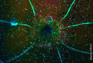
In the new study, scientists from the CECAD Cluster of Excellence for Aging Research, the Center for Molecular Medicine Cologne (CMMC), and the Institute of Genetics (all at the University of Cologne) investigated the function of autophagy in wound healing and in the healthy and uninjured epidermis (skin) of the fruit fly Drosophila melanogaster. In the process studied here, autophagy hence did not digest viruses or bacteria, but the cell's own cell membrane, so that boundaries between cells broke down and a large cell with multiple nuclei was formed.
The team observed that autophagy is increased in the cells surrounding the wound. This leads to a selective breakdown of the membranes that connect the cells. Eventually, a large, multinucleated cell (syncytium) is formed. ‘When we genetically triggered autophagy in healthy, uninjured skin, we again observed the same phenomenon: the membrane between adjacent cells is lost and large patches of multinucleated syncytia form throughout the epidermis,’ said Parisa Kakanj, first author of the study. ‘It is surprising that the formation of syncytia, which we already know to occur in the formation of some organs such as muscles or placenta, also occurs in wound healing. The role of autophagy in this may also be important for our understanding of disease mechanisms, since multinuclear cells are also found in tumors and infected tissues.’
In previous studies of muscle and placenta development, the formation of a syncytium was shown to provide mechanical stability and a strong barrier function to protect tissues from pathogens. Whether the process performs similar functions in wound healing is not yet clear. This will be the subject of future studies.
The investigators also observed an interaction of the autophagic vesicles with the lateral plasma membrane of the cell in the healthy, unwounded epidermis as well as in the cells surrounding the wound. In this context, proper function of the TORC1 protein complex, which has a central regulatory function in cell metabolism, and here also controls autophagy, is crucial to prevent destruction of the epidermis by autophagy. Based on these and other observations, the team hypothesizes that the lateral plasma membrane is a potential source of the autophagic vesicles. ‘Autophagy is a double-edged sword, the precise extent and spatiotemporal activity of which determines whether it is useful or problematic,’ said Maria Leptin. To be able to use the beneficial side of autophagy for prevention and therapy, it is even more important to understand how autophagy is activated and how autophagic vesicles are formed.
Media contact:
Dr. Parisa Kakanj
Institute for Genetics
+49 221 470 3528
pkakanj@uni-koeln.de
Professorin Dr. Maria Leptin
Institute for Genetics
+49 221 470 3401
mleptin@uni-koeln.de
Press and Communications Team:
Dr. Anna Euteneuer
+49 221 478 84043
anna.euteneuer@uni-koeln.de
Publication:
https://www.embopress.org/doi/full/10.15252/embj.2021109992