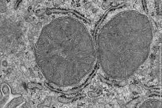
A team of researchers at the University of Cologne and the Max Planck Institute for Metabolism Research has shown what happens in the body when we are hungry and see and smell food. They observed in the mouse model that the liver mitochondria already adapt after a few minutes. Stimulated by the activation of a group of nerve cells in the brain, the mitochondria of the liver cells change and prepare the liver to adapt the sugar metabolism. The results of the study ‘The dynamic state of a prefrontal–hypothalamic–midbrain circuit commands behavioral transitions’ have now been published in Science and could create new opportunities for the treatment of one of the most common illnesses, type 2 diabetes mellitus. The study was led by Professor Dr Jens Brüning, Principal Investigator at the CECAD Cluster of Excellence for Aging Research at the University of Cologne and Director of the Center for Endocrinology, Diabetes and Preventive Medicine at University Hospital Cologne. Professor Brüning is also Director of the Max Planck Institute for Metabolism Research.
Mitochondria in the liver get ready
The researchers gave hungry mice food. However, the mice could only see and smell the food, but not eat it. After just a few minutes, the researchers analysed the mitochondria in the liver and found that processes normally stimulated by food intake were activated.
The findings show that it is sufficient for the mice to see and smell food for a few minutes to influence the mitochondria in the liver cells. This is mediated by a previously uncharacterized phosphorylation in a mitochondrial protein. Phosphorylation describes the reversible attachment of a phosphate group to a protein and is an important modification for the regulation of protein activity. The researchers also show that this phosphorylation affects the sensitivity of the liver to insulin. They have thus discovered a new signalling pathway that regulates insulin sensitivity in the body.
Nerve cells in the hypothalamus
The effect on the liver is mediated by a group of nerve cells called POMC neurons. These neurons are activated within seconds by the sight and smell of food, signalling to the liver to prepare for the incoming nutrients. The results also showed that the activation of POMC neurons alone is sufficient to adapt the mitochondria in the liver even in the absence of food.
“When our senses detect food, our body prepares for food intake by producing saliva and digestive acid. We knew from previous studies that the liver also prepares for food intake,” explained Dr Sinika Henschke, researcher at the Max Planck Institute for Metabolism Research and first author of the study. “Now we took a closer look at the mitochondria in liver cells because they are essential cell organelles for metabolism and energy production, and realized how surprisingly fast this adaptation takes place.”
Principal Investigator Jens Brüning said: “Our results show how closely the sensory perception of food, adaptive processes in the mitochondria and insulin sensitivity are linked. Understanding these mechanisms is also important because insulin sensitivity is impaired in type 2 diabetes mellitus.”
Media Contact:
Professor Dr Jens Brüning
CECAD Cluster of Excellence for Aging Research
Director of the Center for Endocrinology, Diabetes and Preventive Medicine at University Hospital Cologne
Press and Communications Team:
Jan Voelkel
+49 221 470 2356
Publication:
https://doi.org/10.1126/science.adk1005
Further information:
